High-Throughput Differential Scanning Fluorimetry on the Ginkgo Reconfigurable Automation Cart (RAC) Platform

Introduction
Author: Alexander H. Smith
Thermal stability describes a protein’s resistance to conformational change as temperature rises. It is a core developability attribute for therapeutic antibodies: candidates that unfold at low temperature are more prone to aggregation, fragmentation and loss of potency during manufacturing and storage, so thermal stability is measured early and on every molecule that enters a discovery campaign.
Protein thermal stability by extrinsic fluorescence (PTS-EF), widely known as differential scanning fluorimetry (DSF), measures this property using an environmentally-sensitive dye. In its native state a protein is held together by non-covalent interactions. As it is heated, kinetic energy overcomes those interactions, a domain unfolds, and the previously buried hydrophobic surface becomes exposed. Binding of SYPRO™ Orange (Invitrogen S6650) to newly exposed hydrophobic regions triggers an increase in fluorescence, allowing thermal unfolding events to be reported by plotting relative fluorescence units (RFU) against temperature. The temperature at which unfolding begins is the onset temperature (Tonset) and the temperature at which half of a given domain population is unfolded is that domain’s melting temperature (Tm). In an IgG, up to three transitions are typically resolved, corresponding to the CH2, CH3 and Fab domains. Melting transition temperatures are indexed as Tm1-Tm3 [1-3].
PTS-EF is attractive for screening because the reagent cost is low, the readout leverages standard, widely available qPCR hardware, and the assay tolerates crude, partially purified material. When executed on walk-up automation, the method requires an operator to dilute a light-sensitive dye, shuttle plates between an acoustic dispenser, a liquid handler, a sealer and a thermal cycler, and then manually inspect every thermogram to confirm the melting temperatures. Throughput is limited by these requirements as each 384-well plate requires two-three hours of hands-on time.
Here, we describe PTS-EF running end-to-end on Ginkgo’s Reconfigurable Automation Cart (RAC) platform as a fully automated protocol, configured and executed via Ginkgo’s Catalyst Orchestrator software, with data retrieval and analysis driven through the Catalyst API and agentic AI integration. The method was evaluated across 13 fully automated 384-well plates and more than 3,200 assay wells, run in three batches by two operators. Each of these runs passed every qualification criterion and reproduced the walk-up method’s transition counts and melting temperatures with high precision.
Workflow
Fully Automated Workflow
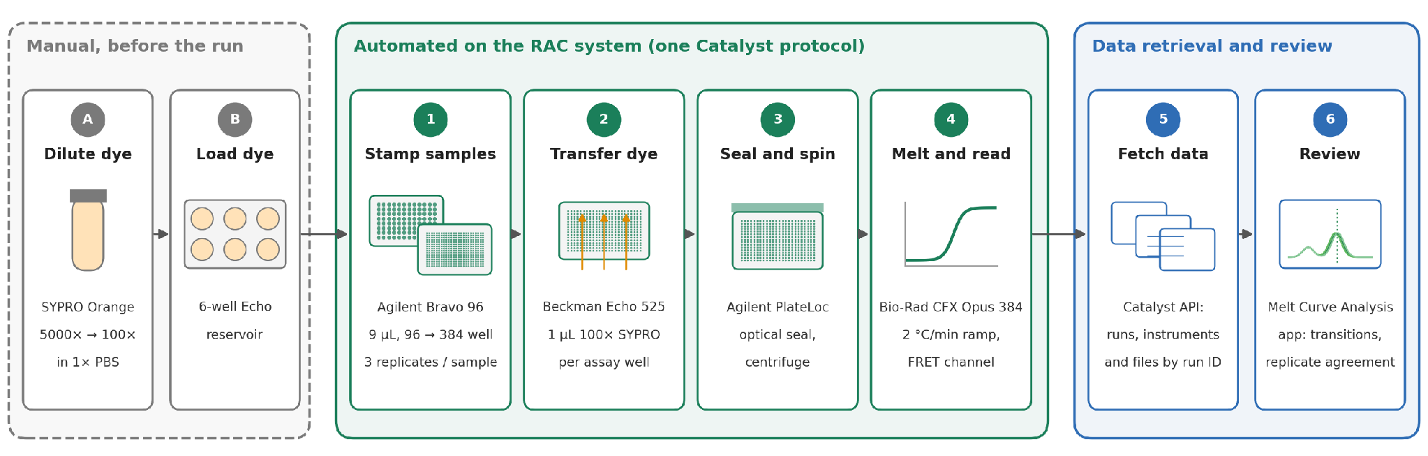
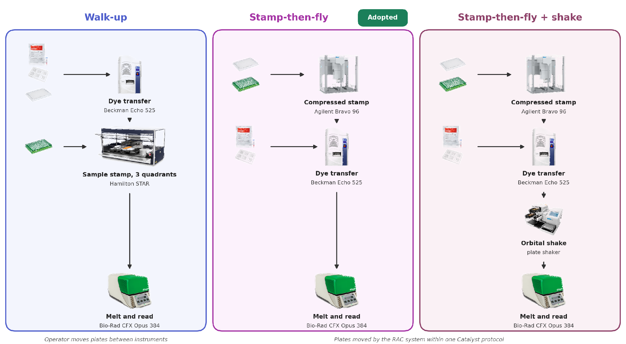
The automated workflow consists of the following main steps (Fig 1), previously executed in a partially automated manner using two benchtop workstations (Beckman Coulter Echo 525 and Hamilton STAR liquid handler) with manual plate transport between them:
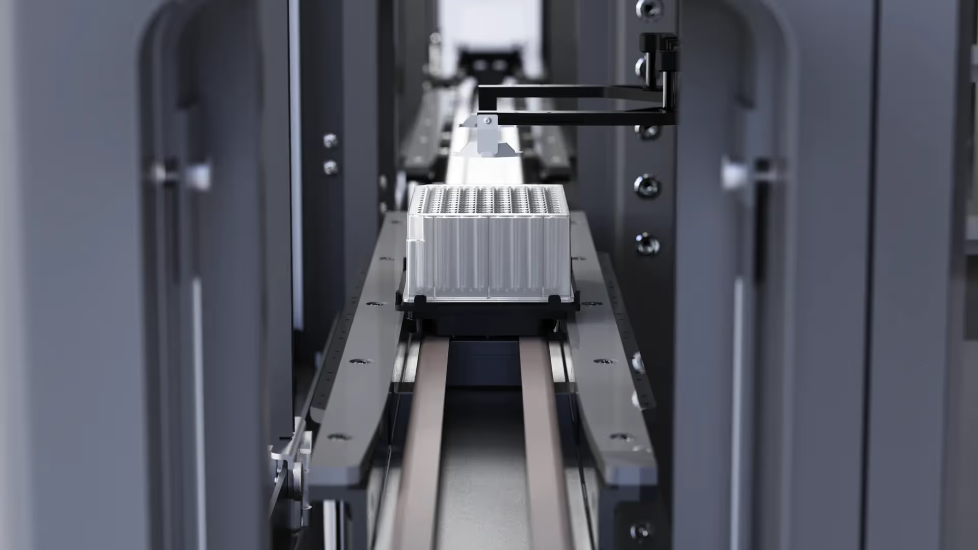
- 96- to 384-well sample replication: 9 µL antibody sample, normalized to 0.25 mg/mL, is stamped from one 96-well input plate (Eppendorf 951020443) into quadrants 1, 2 and 3 of the 384-well assay plate (Eppendorf 951020702) on an Agilent Bravo 96-channel head using a single compressed stamp operation.
- Dye transfer: 1 µL of 100× SYPRO is acoustically transferred from the reservoir to each assay well of the 384-well PCR plate on a Beckman Coulter Echo 525 using a Ginkgo LIMS-generated hitpick map.
- Seal and spin: The assay plate is sealed with an optically clear seal (Agilent PlateLoc thermal microplate sealer; Agilent 16985-001) and centrifuged (BioNex Solutions HiG) to consolidate volume in each well and remove bubbles.
- Extrinsic-fluorescence melt: The plate is loaded into a Bio-Rad CFX Opus 384, which ramps at 2 °C/min while reading in the FRET channel. Source plates and the dye reservoir are re-sealed and returned to temperature-controlled storage.
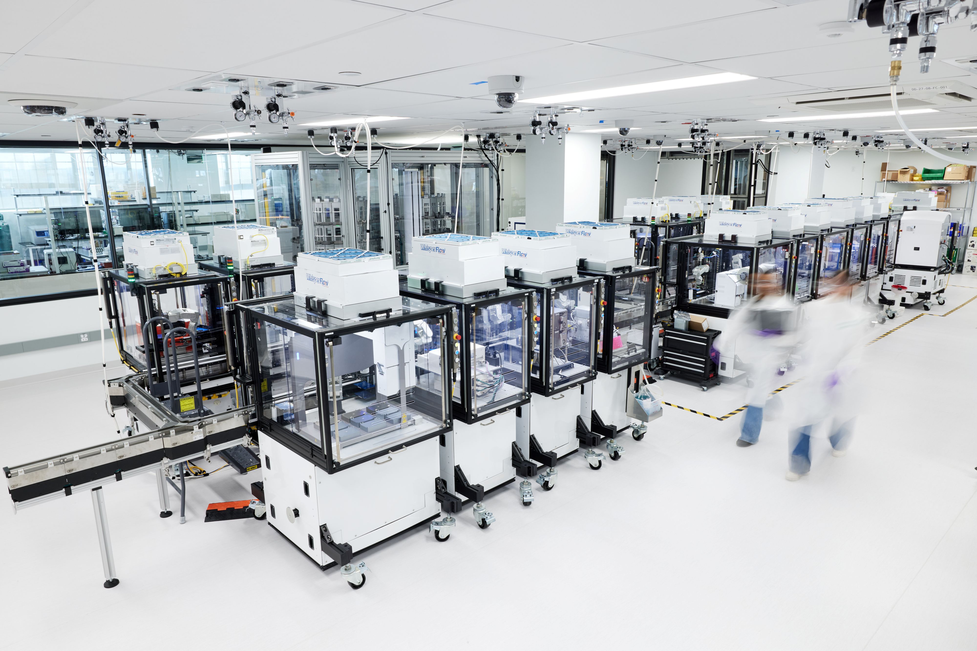
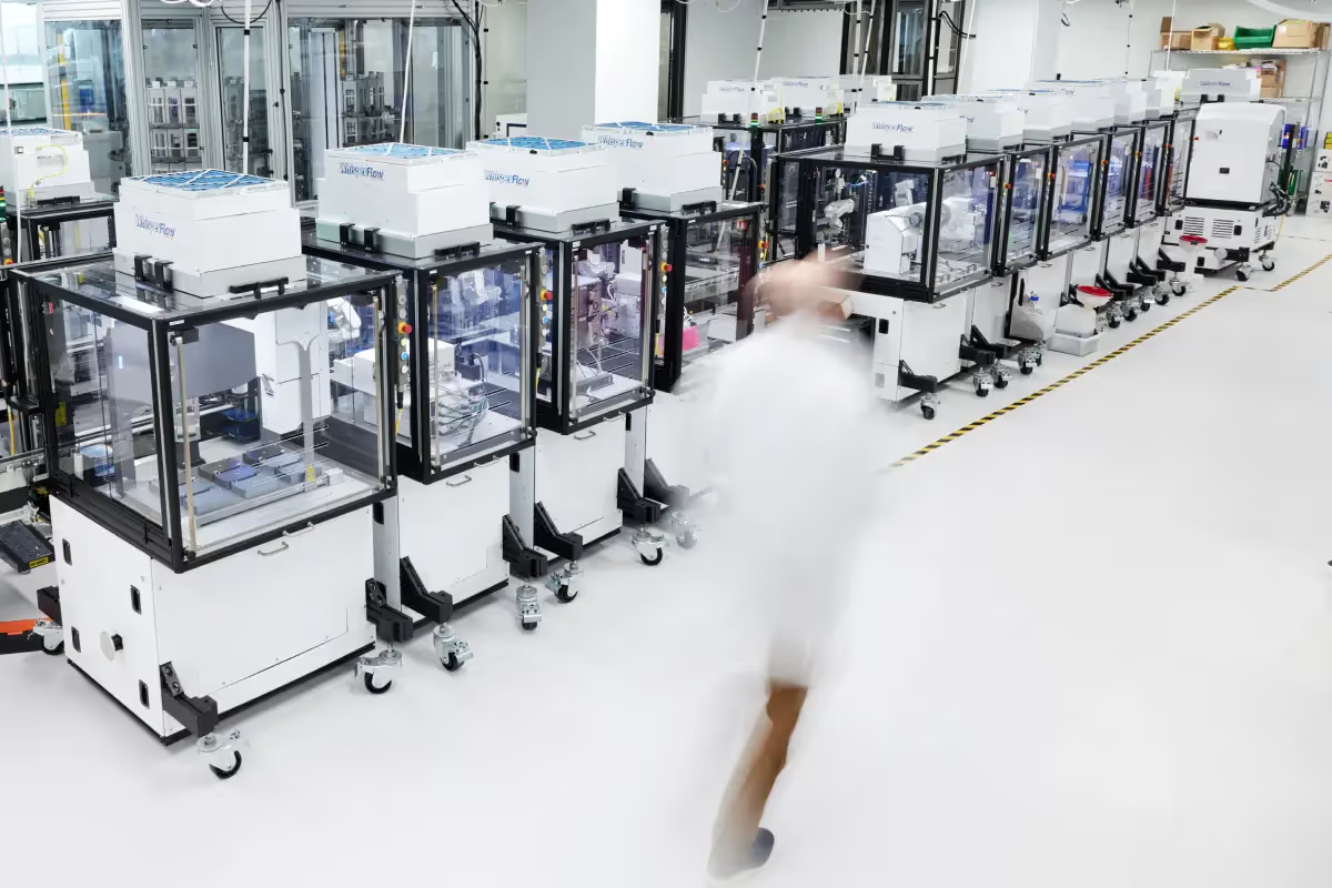
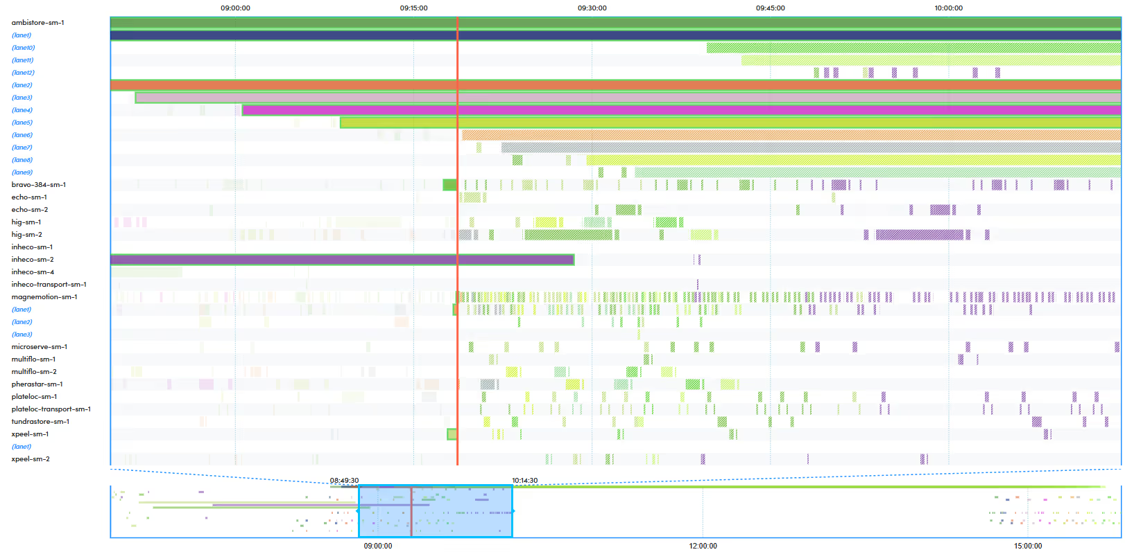
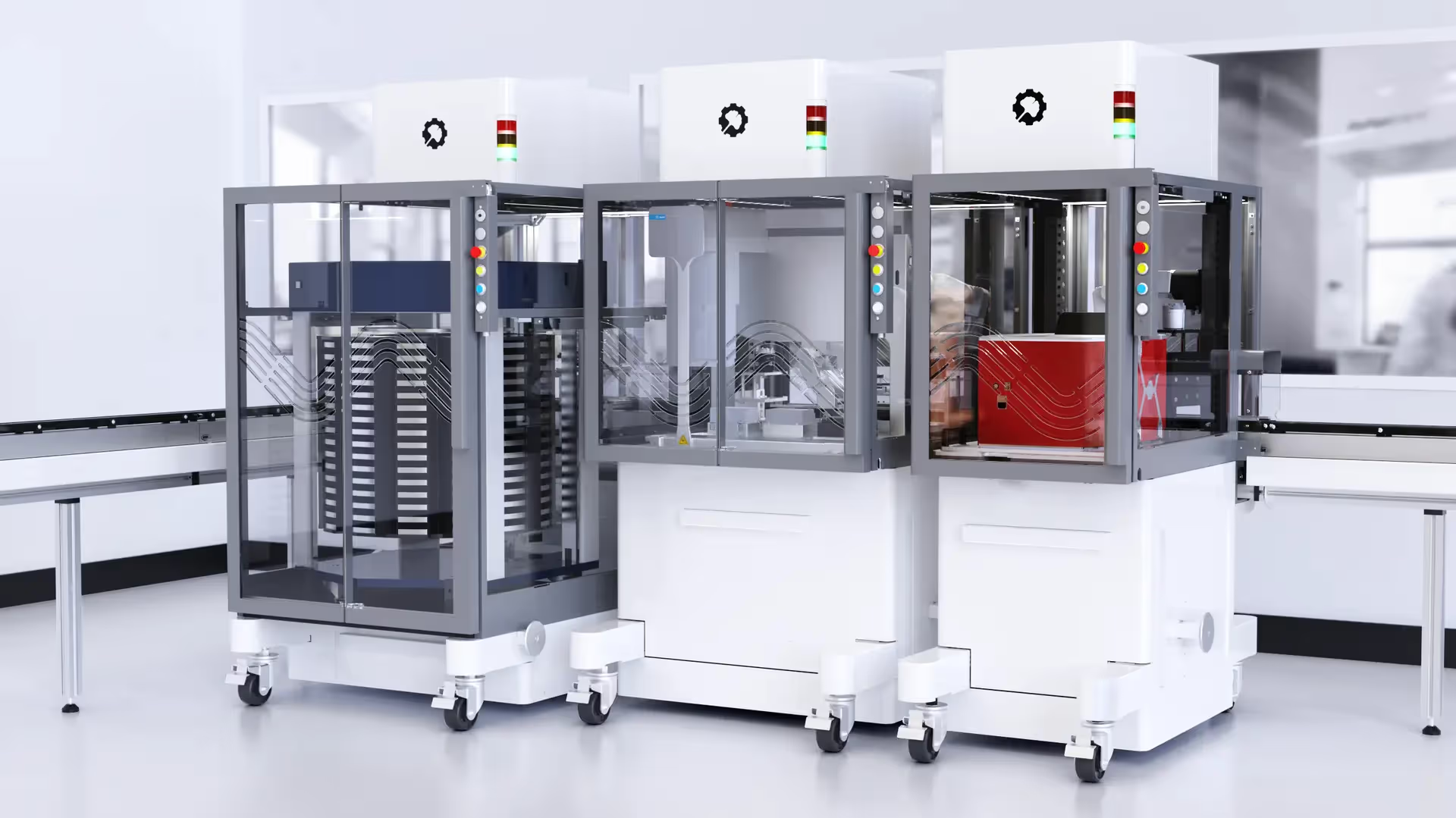
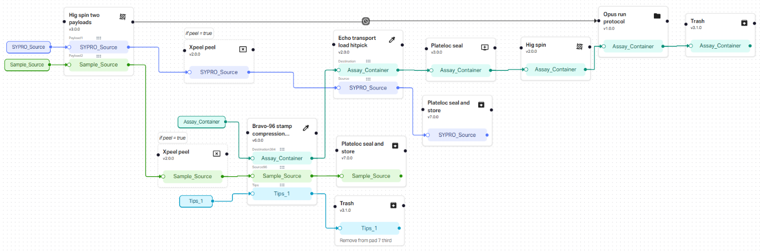
The only manual preparation is diluting SYPRO Orange to its 100× working concentration and loading it into a 6-well Echo reservoir. All subsequent steps are orchestrated via a single Catalyst protocol (abdev_pts-ef_stamp-then-fly) launched via a Catalyst Protocol Launcher web form or agent skill; plate transport between the Echo, Bravo, PlateLoc, HiG centrifuge and CFX Opus is handled by the RAC system’s robotic arms (Mecademic Meca500) and magnetic transport track (Rockwell Automation, MagneMover LITE) with no operator involvement once samples and consumables are loaded (Fig 2). Individual or batched single-plate protocol runs can be flexibly submitted for processing, with Ginkgo’s Catalyst Orchestrator software handling their scheduling and execution, alongside protocol runs submitted by other system users, factoring in critical, user-predefined time constraints and current system load.
Once a run is scheduled, each plate completes in about one hour of active execution and 2 hours of a temperature-ramping thermal cycler protocol with no operator involvement. Ginkgo’s Catalyst software records each plate’s queue time, execution time and step history alongside its experimental data.
Workflow Optimization
Three design choices make the method robust on a shared automation system:
- Stamp-then-fly: A single compressed stamp on the Agilent Bravo 96 replicates each 96-well input plate into three quadrants of the 384-well assay plate in one operation before the Echo adds dye. Compared with sequential quadrant stamping, it uses one tip set instead of three, thus running faster and keeping the assay plate uncovered for less time. This also avoids dye evaporation entirely, since dye is transferred after the protein sample and sealed immediately following. Of the automated method variants tested, this order of operations gave the lowest replicate variability (Fig 3).
- Time constraint addition: A 4,800 second global time constraint restricts how long the assay plate spends unsealed while leaving the scheduler room to place runs efficiently on a busy system. This allows for flexible workflows while mitigating possible sample evaporation or contamination.
- Fluorescence-range QC: Each replicate group is checked for consistent signal and if the raw fluorescence range varies by more than 30% across replicates, the group is flagged. This catches under-filled or evaporated wells that could otherwise return a plausible-looking curve, preventing delivery of untrustworthy data.
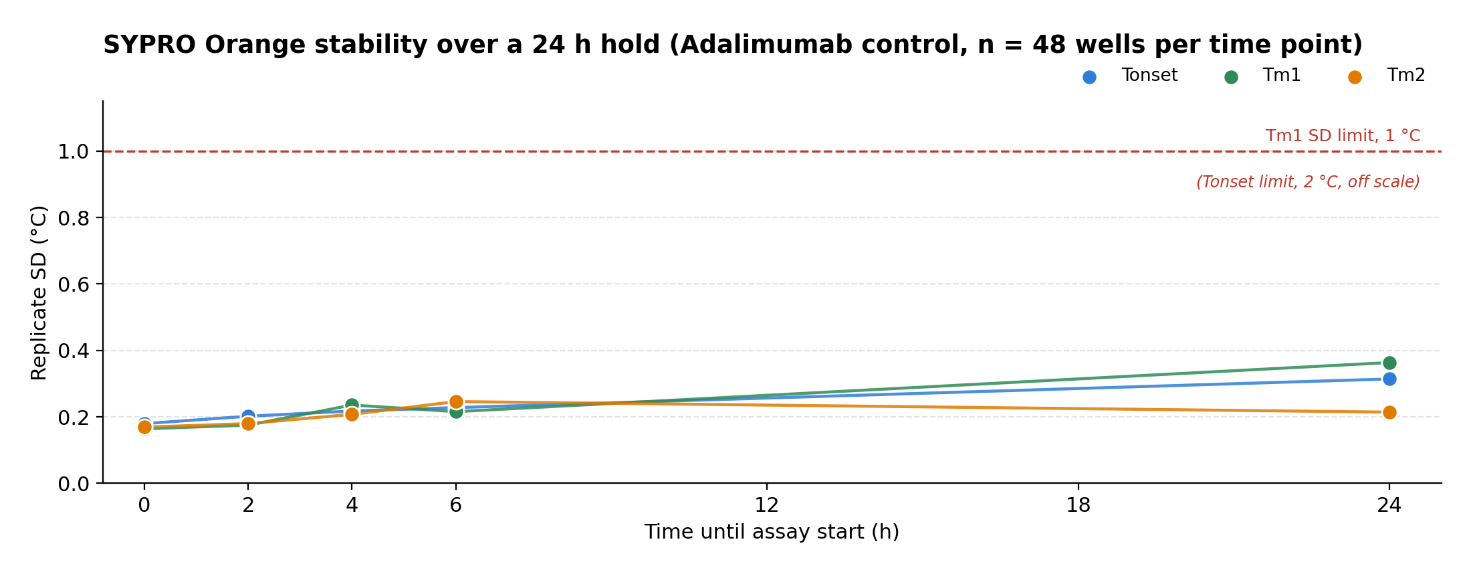
Diluted to its working concentration, SYPRO Orange dye is also robust to typical protocol run queue times. Over a 24 hour hold, standard deviation among the 3 replicates (Fig 4) stayed well inside the assay’s acceptance limits (Table 1), so batches can wait in the queue on a busy, shared system, executing a wide variety of unrelated workflows in parallel, without compromising data quality.
Results
AI-Enabled Data Analysis Through the Catalyst API
Once the bench work is automated, analysis becomes the slowest part of the workflow. Previously, transitioning from raw melt data to an analyzed result required a series of manual steps: locating the specific instrument that the assay was run on, opening the vendor analysis software to export RFU data, and uploading the data to the analysis tool. Thermogram review then took one to two hours per plate.
Ginkgo’s Catalyst exposes protocol run information, instrument assignments and output files through its API, so analysis can start from the automation record rather than from the instruments. Given the time a batch was launched, an AI agent with access to the Catalyst API finds the protocol runs, identifies the instrument each plate ran on and retrieves the data files automatically by run ID. The same query returns queue and execution times, step durations for the thermal-cycler protocol and an error log for each run, so each plate’s processing history arrives with its data.
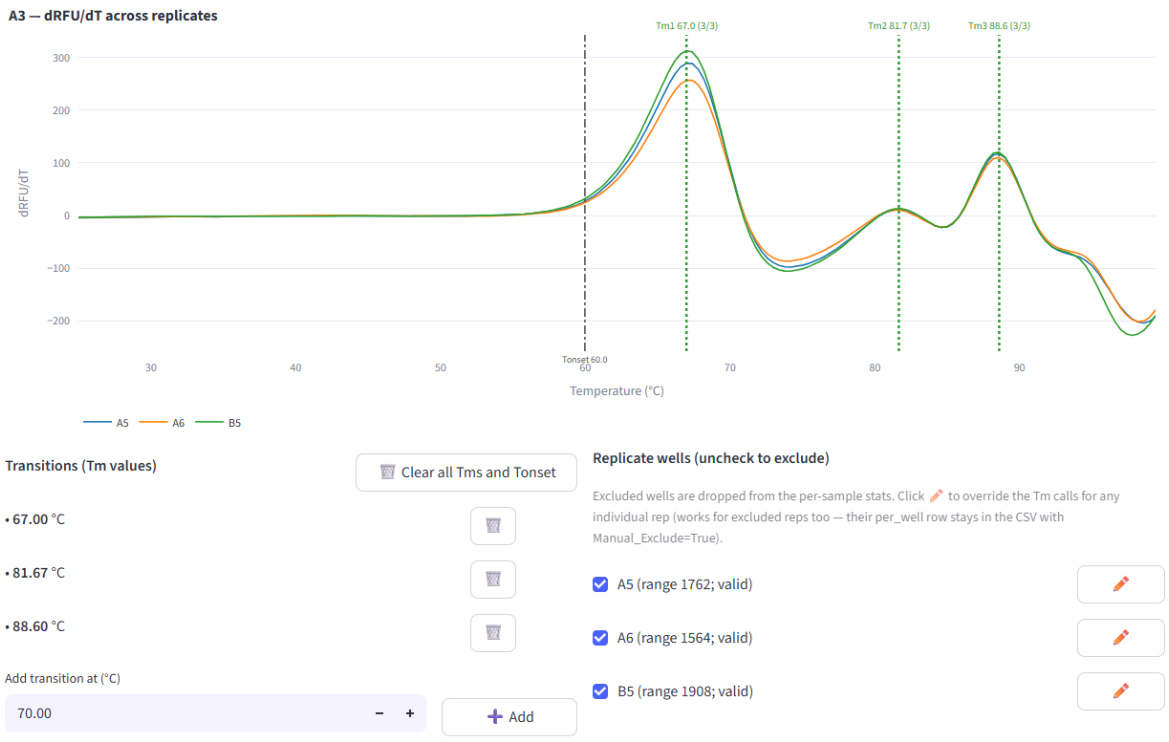
The files then go to a custom-built Melt Curve Analysis application that accepts both .xml and .csv instrument exports, reads sample metadata from the file names, drops empty wells and fits transitions. Replicate thermograms are overlaid for review, with each transition color-coded by how many replicates share it: green for all three, orange for two, red for one. Reviewers can adjust fitting parameters, add or remove transitions, exclude individual replicates or synchronize calls across a replicate group, then export results ready for the downstream dashboard (Fig 5).
Including analysis, total processing time per plate is about half that of the previous workflow. Because run records and results are linked through the API, every plate in the validation below was scored by the same scripted QC checks, with its processing history attached.
Validation Results
Method qualification followed a staged framework, with pass/fail criteria set before any data were collected. Each stage adds scale or a new source of variability:
- The first stage required that a single plate of controls meets the control acceptance criteria: the Adalimumab positive control has a replicate standard deviation below 1 °C for Tm1 and below 2 °C for Tonset, and no buffer blank reports a transition in two or more of its three replicates. Measured transition temperatures are required to align with historical values.
- The second stage required a library plate to meet plate acceptance criteria. One plate of library samples in triplicate, with passing controls, at least 95% of library replicates agreeing on the number of transitions, and a standard deviation below 1 °C across recorded melting temperatures.
- The third stage increased scale by requiring one production-sized (4x 96-well input plates) batch to pass batch acceptance criteria. Every plate must pass the control and sample criteria. At least 95% of samples must agree on the number of transitions within their replicate group, plate and batch, and the standard deviation of sample results between runs must be below 2 °C for at least 95% of samples.
- Production batch results must be replicated at least twice. Passing the same criteria, with one batch executed by a second operator. Similarly to the third stage, each batch contained at least 4x 96-well input plates.
All data were generated on a six-antibody panel (Adalimumab, Bococizumab, Dacetuzumab, Dalotuzumab, Elotuzumab, Ixekizumab) in 43 mM acetate / 148 mM HEPES, pH 6.0, and scored against a manually processed walk-up reference plate.
In three separate batches run on 3 different days, 13 fully automated 384-well plates and more than 3,200 assay wells were processed between two operators and the method passed every qualification criterion (Table 1). Every plate matched the walk-up reference’s transition calls on at least 95% of wells (98.6% pooled; Fig 6), 99.6% of library wells returned usable melting temperatures, and no buffer blank reported a transition.
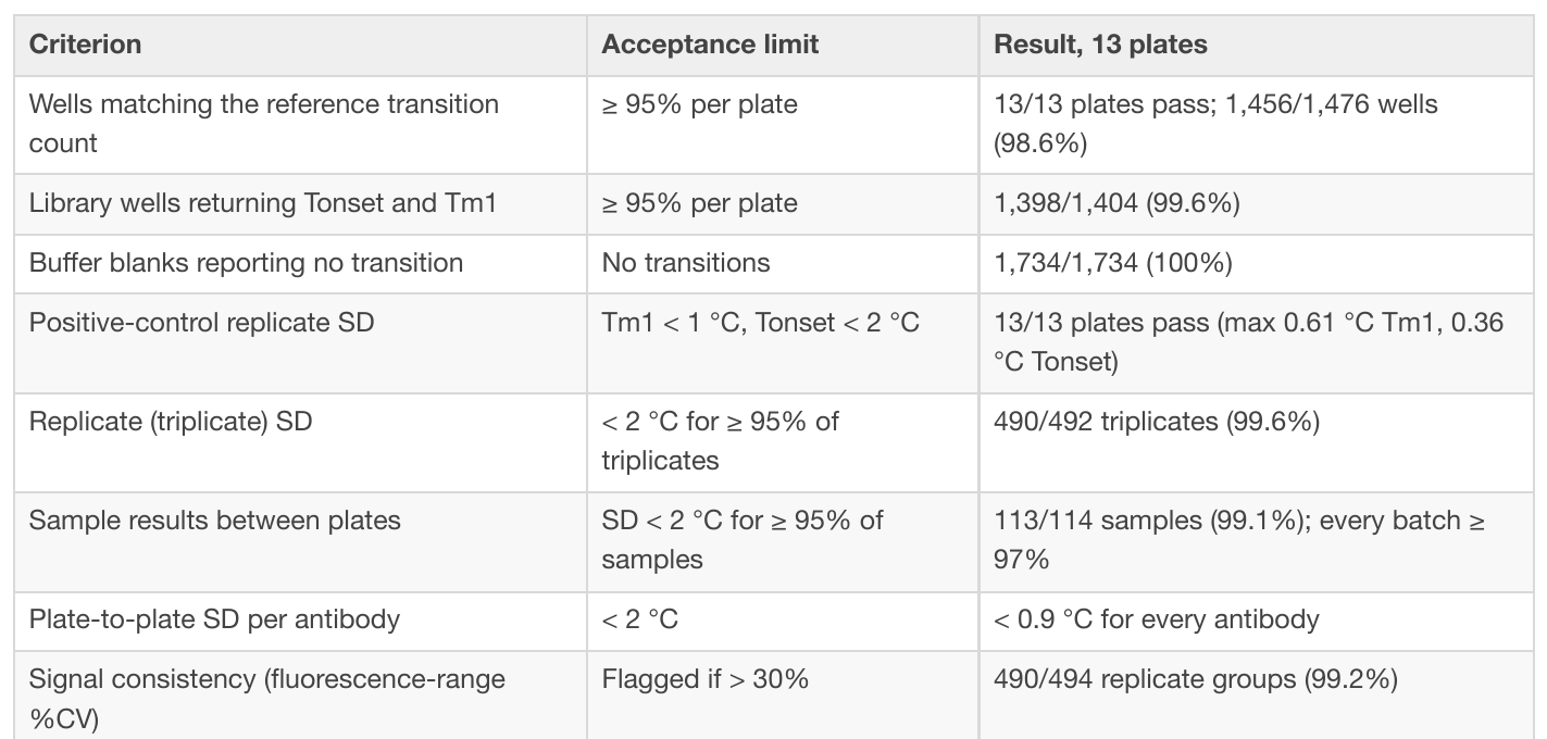
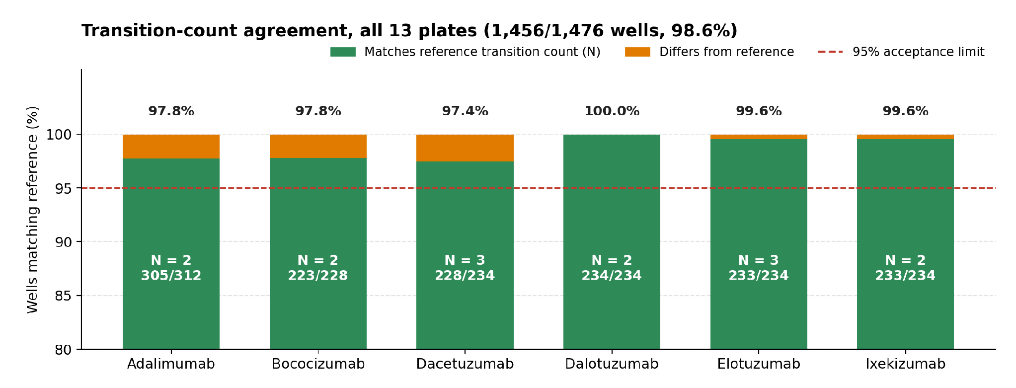
Plate-to-plate reproducibility was high: the standard deviation of per-plate means stayed below 0.9 °C for every antibody, against a 2 °C limit. Automated melting temperatures read slightly warm of the walk-up reference, by +0.5 to +1.3 °C in Tonset depending on the batch. The offset is well inside the acceptance limit and does not affect precision; absolute values should simply be compared with walk-up data with it in mind.
Conclusions
Moving PTS-EF onto Ginkgo’s RAC platform turns a hands-on, multi-instrument assay into a fully automated, walkaway protocol. Replicate stamping, acoustic dye transfer, sealing, centrifugation and the melt read run with no in-person monitoring, and Ginkgo Catalyst software-connected data analysis halves total processing time per plate.
The method passed every qualification criterion across 13 plates and more than 3,200 assay wells, run in three batches by two operators. Every plate matched the walk-up reference’s transition calls on at least 95% of wells, 99.6% of library wells returned usable melting temperatures, no buffer blank reported a transition, and plate-to-plate standard deviation stayed below 0.9 °C for every antibody.
The approach extends beyond the assay execution. Once bench steps run on the RAC platform, the Catalyst API lets an AI agent automatically retrieve run information, instrument assignments and data files directly, so downstream data analysis keeps pace with automated execution and every plate is scored the same way. Adding automated -80°C storage and on-deck reagent dilution would remove the remaining manual step and make the workflow fully drop-off-and-forget.
References
1. Niesen, F.H., Berglund, H. and Vedadi, M. (2007) ‘The use of differential scanning fluorimetry to detect ligand interactions that promote protein stability’, Nature Protocols, 2(9), pp. 2212–2221.
2. Ericsson, U.B., Hallberg, B.M., DeTitta, G.T., Dekker, N. and Nordlund, P. (2006) ‘Thermofluor-based high-throughput stability optimization of proteins for structural studies’, Analytical Biochemistry, 357(2), pp. 289–298.
3. Goldberg, D.S., Bishop, S.M., Shah, A.U. and Sathish, H.A. (2011) ‘Formulation development of therapeutic monoclonal antibodies using high-throughput fluorescence and static light scattering techniques’, Journal of Pharmaceutical Sciences, 100(4), pp. 1306–1315.
Materials
- One (1) to eight (8) 96-well PCR microplates (Eppendorf 951020443) with each well pre-filled with ≥35 µL of normalized antibody sample at ≥0.25 mg/mL (assay limit of detection: 0.15 mg/mL)
- Antibody panel used for qualification: Adalimumab (Ichor Bio ICH4001, 53 mg/mL in PBS; positive control and library member), Bococizumab, Dacetuzumab, Dalotuzumab, Elotuzumab, Ixekizumab
- Buffer blanks (negative control) in the sample background buffer: 43 mM citrate / 148 mM HEPES pH 6.0, 43 mM acetate / 148 mM HEPES pH 6.0, 20 mM HEPES / 150 mM NaCl pH 7.4, or 10 mM histidine / 20 mM NaCl pH 6.0
- SYPRO™ Orange Protein Gel Stain, 5000× in DMSO (Invitrogen S6650), diluted 1:50 in 1× PBS pH 7.5 (Teknova P5275) to a 100× working solution
- One (1) 6-well Echo-qualified reservoir (Beckman Coulter ER-0050) as the acoustic dye source; maximum working volume ~2.5 mL per well
- One (1) 384-well clear PCR microplate per 96-well input (Eppendorf 951020702) as the assay container
- Optically clear microplate seal (Bio-Rad Microseal ‘B’ film MSB1001)
- One (1) rack of 96-well tips per run (single compressed stamp)
Instrumentation: Beckman Coulter Echo 525 acoustic liquid handler; Agilent Bravo 96-channel liquid handler; Agilent PlateLoc thermal microplate sealer; BioNex HiG3 centrifuge; Bio-Rad CFX Opus 384 thermal cycler (FRET channel, 2 °C/min ramp); Ginkgo Nebula RAC system with 4 °C and ambient input storage, orchestrated by Ginkgo Catalyst.
{{data}}
Subscribe to receive future application notes
- 50% Reduction in execution and data analysis time per plate
- <0.9°C Plate-to-plate standard deviation for any antibody, against a 2 °C limit
- ≥99% Of library wells returning viable melting temperatures, in every batch
More Application Notes